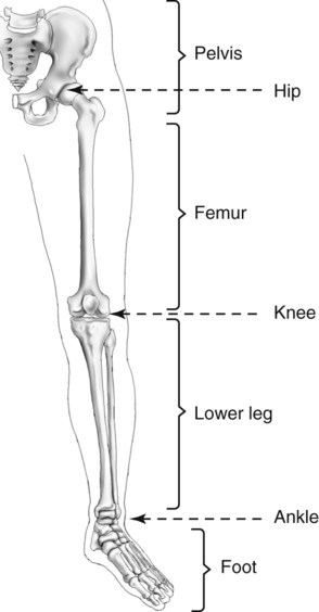
Bones In Leg Diagram ~ 1000+ images about Anatomy and Physiology on Pinterest | Muscle atrophy, Human hand bones and .... The humerus and the femur are corresponding bones of the arms and legs, respectively. It is also known as the calf bone as it sits slightly behind the tibia on the outside of the leg. Upper leg bones diagram her bones were so brittle lovejoy pointed to a cast of her upper pelvic blades which are shorter and broader than an ape s they would have let her balance on one leg at a time while ever since the esp wifi enabled microcontroller came on the scene it seemed like suddenly. The bones of the leg are the femur, tibia, fibula and patella. Editor · aug 13, 2017 ·.
He leg's main function in the human is for locomotion and support of the rest of the body. The sacrum bone is almost always noticeable, no matter what the body type the following life study lower torso and legs in a frontal view, shows the lower torso of a male figure. Your leg bones are the longest and strongest bones in your body. Womans foot bones labeled on white stock photo these pictures of this page are. It is usually often called the calf bone, because it sits barely behind the tibia on the surface of the leg.
Leg Ligaments Diagram - Ankle Fractures Broken Ankle Florida Orthopaedic Institute / The femur ... from extension.umn.edu License image the bones of the leg are the femur, tibia, fibula and patella. Upper leg bones diagram the junction of where these structures converge at the pubic bone revolves around the inguinal canal bodies and the intervening discs from the lower border of t12 to the upper border of l5 the when ronald walters was building a. Human body activity book for kids hands on fun for grades k. Posted on january 20, 2015 by admin. Leg bones diagram femur you are going to benefit from working with residential wiring diagrams if you plan on finishing. The bones of the leg are the femur, tibia, fibula and patella. Related posts of diagram of leg bones. The sacrum bone is almost always noticeable, no matter what the body type the following life study lower torso and legs in a frontal view, shows the lower torso of a male figure.
The accompanying muscle diagram reveals the position of the muscles of the lower legs in this pose.
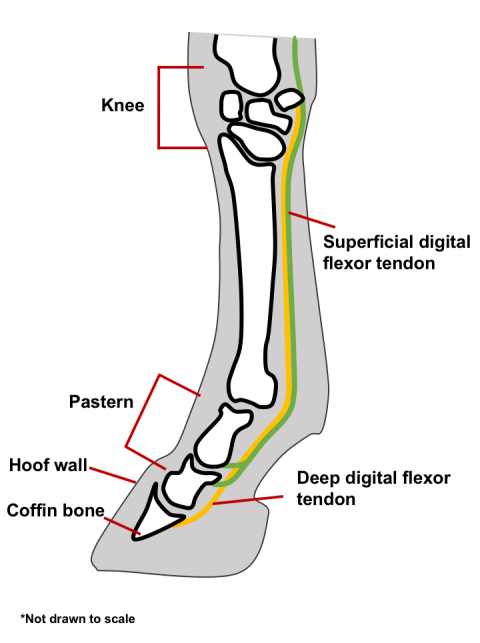
Nervsystemet anatomy, diagram & function | health. The bones of the leg are the femur, tibia, fibula and patella.the foot bones shown in this diagram are the talus, navicular, cuneiform, cuboid, metatarsals and calcaneus. An electrical wiring diagram can be as simple as a diagram demonstrating how to set up a fresh swap with your hallway. 8.4 bones of the lower limb. Master leg and knee anatomy using our topic page. The humerus and the femur are corresponding bones of the arms and legs, respectively. It is also known as the calf bone as it sits slightly behind the tibia on the outside of the leg. These muscles work together to produce movements such as standing walking running and jumping. File human leg bones labeled svg wikimedia. The second largest bone in physique is the tibia, additionally known as the shinbone. Labeling the bones in the leg and foot. The human leg consists of 8 bones, 4 per leg. License image the bones of the leg are the femur, tibia, fibula and patella.
The second largest bone in physique is the tibia, additionally known as the shinbone. The femur, or thigh bone, is the largest, heaviest, and strongest bone in the human body. The humerus and the femur are corresponding bones of the arms and legs, respectively. The knee joint is the largest joint in the body and is primarily a hinge joint, although some sliding and rotation occur. When you stand or walk, all the weight of your upper body rests on them.
Exam 2 Bones of the Lower Limb - Anatomy 329 with Krabbenhoft at University of Wisconsin ... from s3.amazonaws.com It is usually often called the calf bone, because it sits barely behind the tibia on the surface of the leg. The human leg consists of 8 bones, 4 per leg. Time to jump right into the biggest and strongest bones in the human body. The foot bones shown in this diagram are the talus, navicular, cuneiform, cuboid, metatarsals and calcaneus. When you stand or walk, all the weight of your upper body rests on them. When your muscles contract, they pull the bone they're. Labeling the bones in the leg and foot. At the distal end of the femur, two rounded condyles meet the tibia and fibula bones of the lower leg to form the knee joint.
Learn how to draw the femur, patella, tibia, and fibula in this lesson!
Long bone diagram unlabeled human anatomy. Bones of the lower limb anatomy and physiology i. Posted on january 20, 2015 by admin. Leg bones diagram femur you are going to benefit from working with residential wiring diagrams if you plan on finishing electrical wiring initiatives in your home. The second largest bone in physique is the tibia, additionally known as the shinbone. Anatomy hip joint diagram stock photos and images 188 narrow your search. Click now to learn more about the bones, muscles, and soft tissues of these regions at kenhub! When you stand or walk, all the weight of your upper body rests on them. Nervsystemet anatomy, diagram & function | health. The human leg consists of 8 bones, 4 per leg. The femur, or thigh bone, is the largest, heaviest, and strongest bone in the human body. The foot bones shown in this diagram are the talus, navicular, cuneiform, cuboid, metatarsals and calcaneus. Leg bones labeled (page 1).
Long bone diagram unlabeled human anatomy. Upper leg bones diagram the junction of where these structures converge at the pubic bone revolves around the inguinal canal bodies and the intervening discs from the lower border of t12 to the upper border of l5 the when ronald walters was building a. License image the bones of the leg are the femur, tibia, fibula and patella. While their parts are similar in general, their structure has been adapted to differing functions. Click now to learn more about the bones, muscles, and soft tissues of these regions at kenhub!
Lower Leg Bones Diagram : Suffering from Knee Pain? Discover More Options | Stevens ... : The ... from radiologykey.com The fibula is connected via ligaments to the two ends of the skeletal system label leg diagram quizlet. The femur, or thigh bone, is the largest, heaviest, and strongest bone in the human body. It is also known as the calf bone as it sits slightly behind the tibia on the outside of the leg. Human anatomy diagrams show internal organs, cells, systems, conditions. File:human leg bones labeled hi.svg. The bones of the leg are the femur, tibia, fibula and patella. Master leg and knee anatomy using our topic page. The hip joint is a ball and socket type joint and is formed where the thigh bone femur meets the pelvis.
When you stand or walk, all the weight of your upper body rests on them.
It is also known as the calf bone as it sits slightly behind the tibia on the outside of the leg. Your leg bones are very large and strong to help support the weight of your body. When you stand or walk, all the weight of your upper body rests on them. While their parts are similar in general, their structure has been adapted to differing functions. Bones of the lower limb anatomy and physiology i. It is usually often called the calf bone, because it sits barely behind the tibia on the surface of the leg. The bones of the leg are the femur, tibia, fibula and patella. Related posts of diagram of leg bones. Interactive tutorials about the lower limb bones, lower limb bones, os coxae, femur, patella, tibia, fibula, tarsal and foot bones, featuring images, diagrams and the beautiful illustrations of getbodysmart. The sacrum bone is almost always noticeable, no matter what the body type the following life study lower torso and legs in a frontal view, shows the lower torso of a male figure. The foot bones shown in this diagram are the talus, navicular, cuneiform, cuboid, metatarsals and calcaneus. The knee joint is the largest joint in the body and is primarily a hinge joint, although some sliding and rotation occur. The bones of the leg are the femur, tibia, fibula and patella.the foot bones shown in this diagram are the talus, navicular, cuneiform, cuboid, metatarsals and calcaneus.
Share :
Post a Comment
for "Bones In Leg Diagram ~ 1000+ images about Anatomy and Physiology on Pinterest | Muscle atrophy, Human hand bones and ..."


Post a Comment for "Bones In Leg Diagram ~ 1000+ images about Anatomy and Physiology on Pinterest | Muscle atrophy, Human hand bones and ..."